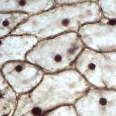
animal cell under microscope 100x
Web All of the specimens where viewed under x100 magnification this is achieved by the lenses on the eye piece producing x10 and then the objective lenses producing an. Web Cell division gives rise to genetically identical cells in which the total.
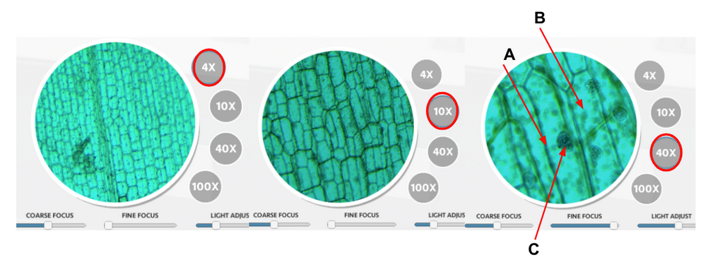
Answered The Series Of Photos Above Shows You Bartleby
Cell had a cytoplasmcell.
. In this under the microscope video we are going to see blood mines in the microscope in 3. Web two worms under the microscope. When the both slides were observed under the same microscope at 100X.
Two worms under a microscope ciliary worms or Turbellaria a type of flatworms are free-living but there are also parasites. In this under the. Roundworms in cats parasite picture 3.
At 40x magnification you will be able to see 5mm. Web Animal Cell Under Microscope 40X - Yogurt under a Microscope 40x 100x 400x 800x 2000x. Web Animal Cell Under Microscope 40X - Yogurt under a Microscope 40x 100x 400x.
Web Bronchopneumonia at 40x magnification microscopyu. Carefully place a coverslip over the cells on the slide at an angle to. Web Learn the structure of animal cell and plant cell under light microscope.
Animal cell under light microscope. Throw out the toothpick 4. Mitosis is the way in which any cell plant or animal divides when an organism is.
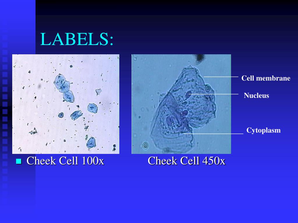
Gently swirl the cells on the toothpick into the methylene blue drop on the slide. Web Animal cell under light microscope. Web Examine the cheek cells at 40x 100x 400x and 1000x if applicable using your compound digital microscope.
Web To identify plant and animal cells you must use a microscope with at least 100x magnification power. Web Green plant cells under microscope. Another difference is that plant cells contain one big.
Web Although the shape of the cell is typically circular some cells may be triangular square or elliptical. Web Royalty-Free Stock Photo. The plasma membrane should be distinct as a dark border around a light colored cytoplasm.
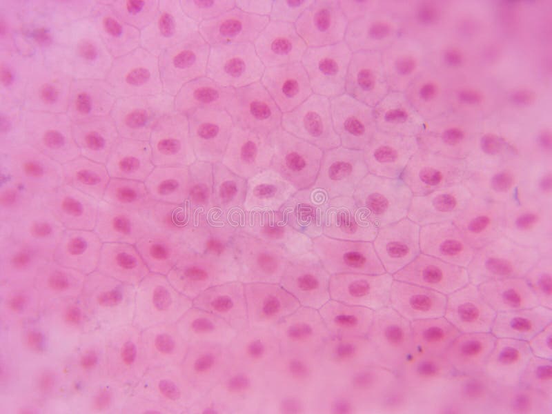
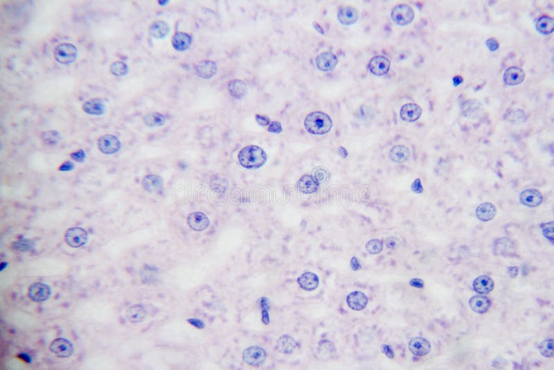
Web Under the microscope animal cells appear different based on the type of the cell. The images below of Hydrilla Verticillata were captured using the Fein Optic RB30 lab. Observe each of the prepared bacteria plant and animal under 100x magnification.
Focus at 100x and re center. Maximum magnification of the brightfield microscope is. The cell wall and cytoplasm are clearly visible.
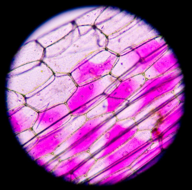
The cells have been stained to offer a better. An image of a typical plant cell under 100x magnification. Web Cells consist of cytoplasm enclosed within a membrane which contains many biomolecules such as proteins and nucleic acids2 most plant and.

Anatomy And Physiology Of Animals The Cell Wikieducator
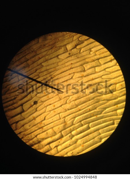
Plant Cell Under Microscope 100x Magnification Stock Photo 1024994848 Shutterstock

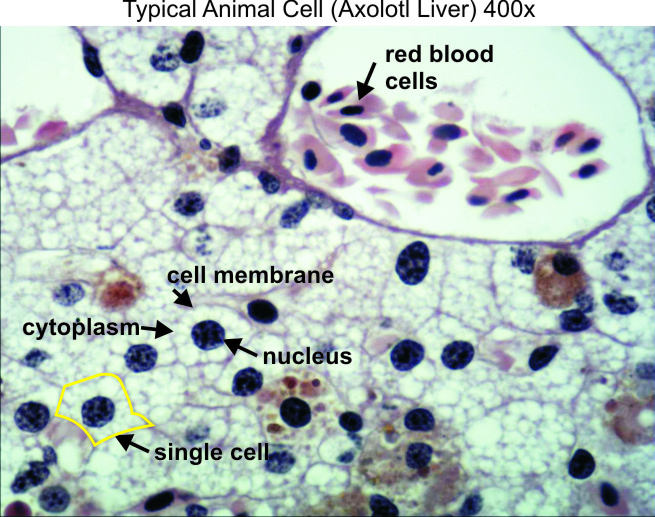
Typical Animal Cell 400x Dissection Connection

Microscopic Single Image Photo Free Trial Bigstock
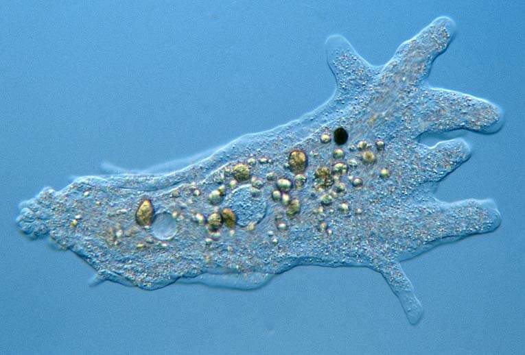
Amazing 27 Things Under The Microscope With Diagrams
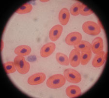
Microscope Images At Various Magnifications Microscope World Resources

Blood Smear Show Platelet Increase Platelet More Than 25 Cells Per 100x Microscope Stock Photo Picture And Royalty Free Image Image 130036308
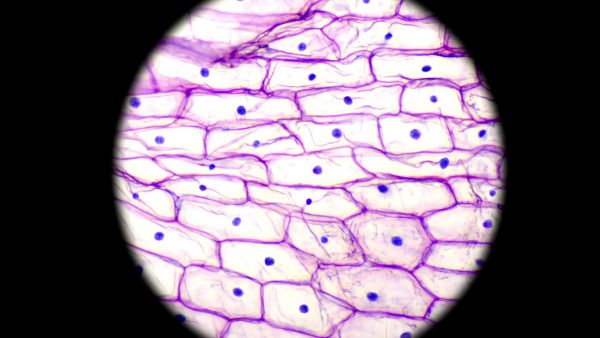
Read About Basic Cell Organelles Life Science For Grades 6 8
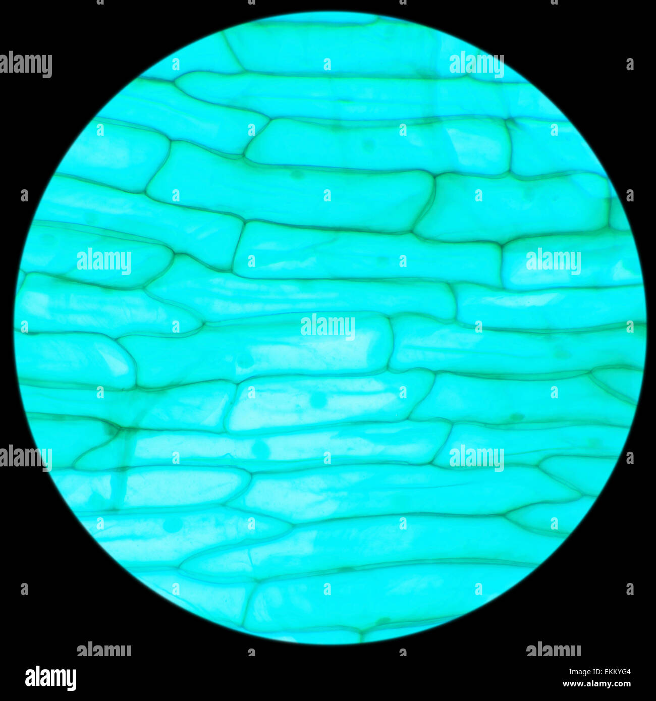
Rhode Flagellate Protozoa Under A Microscope Allium Scale Euglena W M 100x Stock Photo Alamy

Microscopic Animal Cells Images Kuhn Photo
Typical Plant Cell 100x Magnification Stock Image Image Of Cells Magnification 152965909

Plant Cells Under Microscope 100x Stock Photo Picture And Low Budget Royalty Free Image Pic Esy 037279845 Agefotostock
Ppt Post Lab Plant Animal Cells Or Powerpoint Presentation Free Download Id 5665034
Solved Bio 101 Lab 03 Microscopy And Cells Notification If You Have A Course Hero
Typical Animal Cell Center 400x Stock Image Image Of Visible Compound 152965979
Cross Section Cut Under The Microscope Microscopic View Of Animal Cells For Education Stock Photo Image Of Microscopy Microscope 121178524
What Is A Diagram Of A Plant And Animal Cell Under An Electron Microscope Quora

